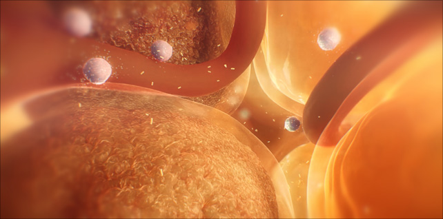
Fat cell enlargement & type 2 diabetes
Apr 22, 2022Fat cell enlargement in obesity is related to insulin resistance and type 2 diabetes.
In this sequence, we travel into the visceral fat and see an active environment – teeming with overstressed, damaged fat cells, vasculature, and immune cells.
Stress on these hypertrophic adipocytes results in lipid spillover (fatty acid release) into the circulation, as well as proinflammatory cytokine release and immune cell recruitment.
Tagged: adipocyte animation, adipocytes, adipose tissue disfunction, diabetes, diabetes 2, diabetes animation, diabetes prevention, fat cell animation, fat cell enlargement, fat cells, hybrid medical, hypertrophic adipocytes, hypertrophic adipose expansion, hypertrophic fat cells, medical animation, medical illustration, medical visualisation, scientific animation, type 2, type 2 diabetes
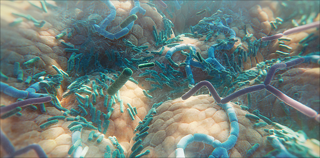
The diverse human gut microbiome
Jun 06, 2023Animation sequence featuring a fly-over of the human gut microbiome – a huge, diverse range of many different types of bacteria residing inside our small and large intestine.
This sequence was created for an episode (for WebMD) that examines how the gut microbiome may influence cancer response to immune checkpoint inhibitors. Essentially, if a patient has melanoma, the types of bacteria in their gut may help determine if the cancer responds to checkpoint inhibitor therapy or not.
There are trillions of microbes in our gastrointestinal tract, around 90% of which are bacteria.
Our microbiome has a profound impact on our well-being and can affect us in several different ways – both beneficial and harmful. In recent years, scientists and researchers have discovered correlations between the composition of an individual’s microbiome and their wider health, including obesity, mental health, bowel disease, and the immune system.
Further advances need to be made in learning more about actual causation – and which effects come from which individual microbes.
Tagged: bacteria, biology, biomedical communications, biotech, biotechnology, gastrointestinal tract, gut bacteria, gut diversity, gut health, gut microbes, gut microbiota, human gut microbiome, hybrid medical, medical animation, Medical science, medical visualization, medicine, microbiology, microbiome, microbiome health, microbiome medicine, pharma, science, science visuals, scientific animation, visual science
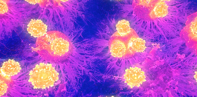
Cytotoxic T cells targeting tumor cells | immunotherapy animation
May 03, 2022A close-up fly-through of tumor cells, and the arrival of cytotoxic t cells intent on eradicating them. Activation and proliferation of cytotoxic T cells are critical for immune-mediated tumor destruction.
A key challenge we were given for this sequence: develop a rendering approach that mimics infrared thermal microscopy imaging techniques.
Researchers have been making advancements in understanding the thermal behavior of cells – for example, the ability to read the signatures of tumor cells due to their higher metabolism – in order to be used alongside existing histological analysis methods.
Tagged: biology, biomedical communications, biotechnology, cancer cells, cytotoxic T cells, fluorescence microscopy, hybrid medical, immune cells, immunology, immunotherapy, infrared, infrared (IR) microscopy, infrared imaging in biology, infrared spectroscopy, IR imaging, IR microscopy, Killer T cell animation, Killer T cells, medical animation, microspectroscopic animation, Minneapolis Minnesota, oncology, pharma, science visuals, scientific animation, T cells, thermal, thermal imaging, thermal microscopy, tumor cells, tumor death
Oligomers, fibrils, & plaques | Alzheimer’s | medical animation
Aug 21, 2023Segment from 5-minute mechanism of action animation.
This sequence explores a hallmark and pathophysiological feature of Alzheimer’s disease: toxic amyloid beta aggregates (Aβ) that accumulate into plaques (neurofibrillary tangles) near neurons in the brain.
A challenge for this sequence was to show the assembly of these naturally occurring peptides – oligomers, fibrils, and plaques – all in one continuous shot.
In addition, we show how over time, extracellular amyloid beta aggregates, including oligomers, fibrils, and plaques, are thought to exert toxic effects on neurons and synapses, disrupt cell function, and lead to synaptic dysfunction and loss.
Tagged: aggregates, alzheimer’s, alzheimer's disease, alzheimers, amyloid, amyloid beta, amyloid beta plaques, amyloid fibril, amyloid plaques, biomedical communications, biotech, brain deterioration, cognitive decline, digital health, extracellular amyloid beta aggregates, fibrils, hybrid medical, medart, medical animation, medical visualization, neurofibrillary tangles, neuroscience, oligomers, plaques, scicomm, synaptic dysfunction, toxic aggregates
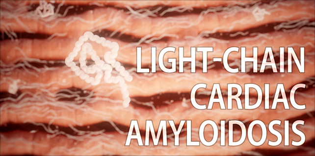
Amyloid light chain (AL) amyloidosis | cardiac amyloidosis animation
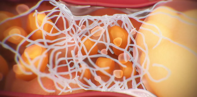
Jul 21, 2023Opening with a view of the heart, we reveal several amyloid deposits infiltrating the muscle tissue.
Then we zoom down to the heart muscle cells and see misfolded light chains aggregating into insoluble amyloid fibrils.
We fade, over time, from the early onset of light-chain infiltration to late-stage fibril aggregation, where the buildup of amyloid deposits around and in between the cardiomyocytes has made the muscle tissue stiff and less flexible, impairing the heart’s ability to relax and fill with blood.
Amyloid light chain (AL) amyloidosis is the most common form of systemic amyloidosis and is caused by the misfolding of immunoglobulin light chains, which are produced by abnormal plasma cells in the bone marrow.
Deposits can be widespread, affecting multiple organs, or they can be more localized.
Tagged: AL amyloidosis, amyloid deposits, amyloid light chain, amyloidosis, cardiac, cardiac amyloidosis, cardiology, fibrils, heart, heart failure, heart health, hybrid medical, immunoglobulin light chains, light chain amyloidosis, medical animation, misfolded proteins, misfolding, myocardium, scientific animation, visual science
Non-alcoholic steatohepatitis (NASH) | NASH liver disease animation
May 16, 2022A few selected sequences we produced for a recent Medscape CME / ABIM MOC / CE symposium titled NASH 101: What You Need to Know Now
The full 2.5- minute animation was developed to be played as a visual aid when the moderator discussed epidemiology and pathogenesis.
We were asked to conceptualize and develop this visual science story. It provides insight into the pathophysiology of NAFLD and NASH, including the role of fatty acids, insulin resistance, and steatosis, as well as mitochondrial dysfunction, excess ROS production, and the stress put on the endoplasmic reticulum inside ballooning hepatocytes.
In addition, we depicted key attributes of the progression of non-alcoholic steatohepatitis (NASH) including bloated hepatocytes and the fat droplets accumulating inside, displaced cell organelles, lobular inflammation, and the scarring (fibrosis) that occurs as collagen fibers fill the spaces once cell death occurs.
In the last few years, nonalcoholic fatty liver disease (NAFLD) and nonalcoholic steatohepatitis (NASH) have become the most common causes of chronic liver disease worldwide. This epidemic is starting to parallel the epidemics of obesity and type 2 diabetes.
Tagged: biology, biomedical communications, cirrhosis, fatty acids, fatty liver disease animation, hepatocytes, hybrid medical, insulin resistance, lipotoxicity, liver animation, medical animation, medical marketing, NAFLD, NASH, NASH liver disease animation, non-alcoholic steatohepatitis, nonalcoholic fatty liver disease, obesity, science visuals, scientific animation, steatohepatitis, steatosis, type 2 diabetes
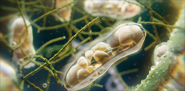
Paramecia: unicellular ciliates animation
Jun 02, 2023A view of paramecia spiraling their way about in a freshwater pond.
In this sequence, we get a close-up look at one of these unicellular microorganisms as it makes its way around using the tiny hair-like cilia on its outer surface. Arranged in longitudinal rows, there are around 4,000 of these motile cilia on its surface, beating in waves to ensure movement and feeding.
In addition to other organelles, we can see several vacuoles inside, digesting organisms such as bacteria and yeasts.
The paramecium was among the first single-celled organisms to be observed under the microscope.
In order to capture the emergent behavior and movement of this microworld, the definitive goal here was to create a natural environment using physics simulations and a procedural pipeline instead of hand-keyed animation.
Tagged: biology, cell, ciliate, ciliates, houdini, houdinifx, hybrid medical, medical animation, micro, microbes, microbiology, microcosmos, microscope, microscope photography, microscopy, microworld, paramecia, paramecium, pharma, photomicrography, redshift, redshift3d, science, science visuals, scientific animation, sidefx, unicellular
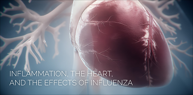
Inflammation, the Heart, and the Effects of Influenza
Mar 15, 2022Sequence from an animation that explores influenza, the body’s immune response to the virus, and how it may cause dangerous levels of inflammation throughout multiple organs systems of the body.
In this excerpt, we depict a plaque rupture and show the important role inflammation plays in the development of atherosclerotic plaques.
This is a sequence from a three-part production that provides insight into how older adults with existing conditions such as cardiovascular disease, respiratory illness, or diabetes may face a higher risk of severe complications when they are infected with the influenza virus.
Tagged: atherosclerosis, cardiac, cardiac arrest, cardiology, cardiovascular disease, flu, heart disease, heart disease awareness, heart failure, hybrid medical, hypertension, immune response, inflammation, influenza, medical animation, plaque, scientific animation, vaccines, virus, visual science
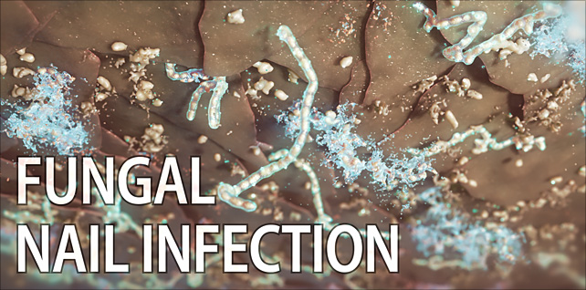
Onychomycosis & nail fungal disease
May 22, 2022This animation takes a closer look at a fungal infection of the toenail and the key attributes of its invasion, including discoloration and brittleness of the nail, striations and thickness of the nail plate, and ultimately, the presence and proliferation of fungi at the nail bed.
One of the more important aspects for this project was to create a highly detailed, diseased nail cross-section, including anatomically correct layers (dorsal, intermediate, and ventral). This would allow us to feature the delivery vehicle and transport of cosmetic and antifungal ingredients through the nail plate.
Additional key challenges for this piece included creating a 3D toe with diseased nail, particle simulations of the key anti-fungal solution ingredients and their pathway down to the nail bed, and the destruction of fungal species (including yeasts, dermatophyte molds, and non-dermatophyte molds) at the nail bed.
Trichophyton rubrum – the fungal species we see at the end – is the most common type of nail fungus.
Tagged: antifungal, cosmetic, dermatology, dermatophyte, fungal, fungal infection, fungal nails, fungus, houdini, hybrid medical, keratin matrix, medical animation, mold, nail bed, nail fungal disease, nail plate, nails, onychomycosis, pedicure, redshift, science, science visuals, scientific animation, toe fungus, toe fungus animation, toe fungus3D, toenail fungus, trichophyton, trichophyton rubrum, yeast
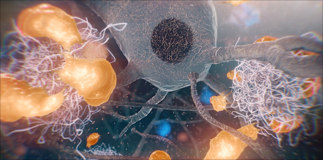
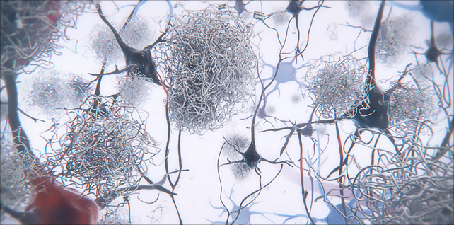
Proliferation and activation of microglia in the brain
Aug 31, 2023Amyloid plaques are the hallmark of Alzheimer’s disease (AD).
This sequence shows the proliferation and activation of microglia in the brain, concentrating around and engulfing neurotoxic amyloid-beta plaques that have caused damage to impaired neurons nearby.
Microglia are specialized immune cells of the central nervous system, specifically in the brain and spinal cord. They play a vital role in monitoring and maintaining the neural environment. One of their primary responsibilities is to detect and remove damaged neurons, plaques, and other cellular debris from the brain.
We worked closely with our clients to create an accurate portrayal of microglia, including their form and structure, their movement and behavior, as well as the manner in which they form a barrier around these growing, toxic plaques. Microglia size, movement, and morphology were all examined by viewing time-lapse imaging projection software – and then recreated in 3D.
One potentially harmful aspect of this relationship between microglia and plaques is that prolonged activation of microglia in response to persistent plaques can lead to chronic inflammation, which might contribute to neuronal damage and the progression of Alzheimer’s disease.
Tagged: aggregates, Alzheimer’s disease, alzheimers, amyloid, amyloid beta, amyloid fibril, amyloid plaques, amyloid-beta plaques, axonal dystrophy, Aβ plaques in Alzheimer’s disease, biomedical communications, biotech, biotechnology, brain deterioration, cognitive decline, glia, hybrid medical, medical animation, medical visualization, microglia, microglia barrier, microglial, microglial phagocytosis, Minneapolis Minnesota, nervous system, neurofibrillary tangles, neurons, neuroprotection, neurotoxic, plaques, scientific animation, synaptic dysfunction, toxic aggregates